
Skin Cancer Treatment
in Thousand Oaks, CA
What Is Skin Cancer?
Skin cancer is the uncontrolled overgrowth of abnormal skin cells. This abnormal growth of cells is caused by unrepaired DNA damage, which triggers a mutation that causes skin cells to multiply quickly, forming cancer cells. Anyone can get this type of cancer, and it can occur anywhere on the body, but it’s more likely to develop when your skin is regularly exposed to sunlight.
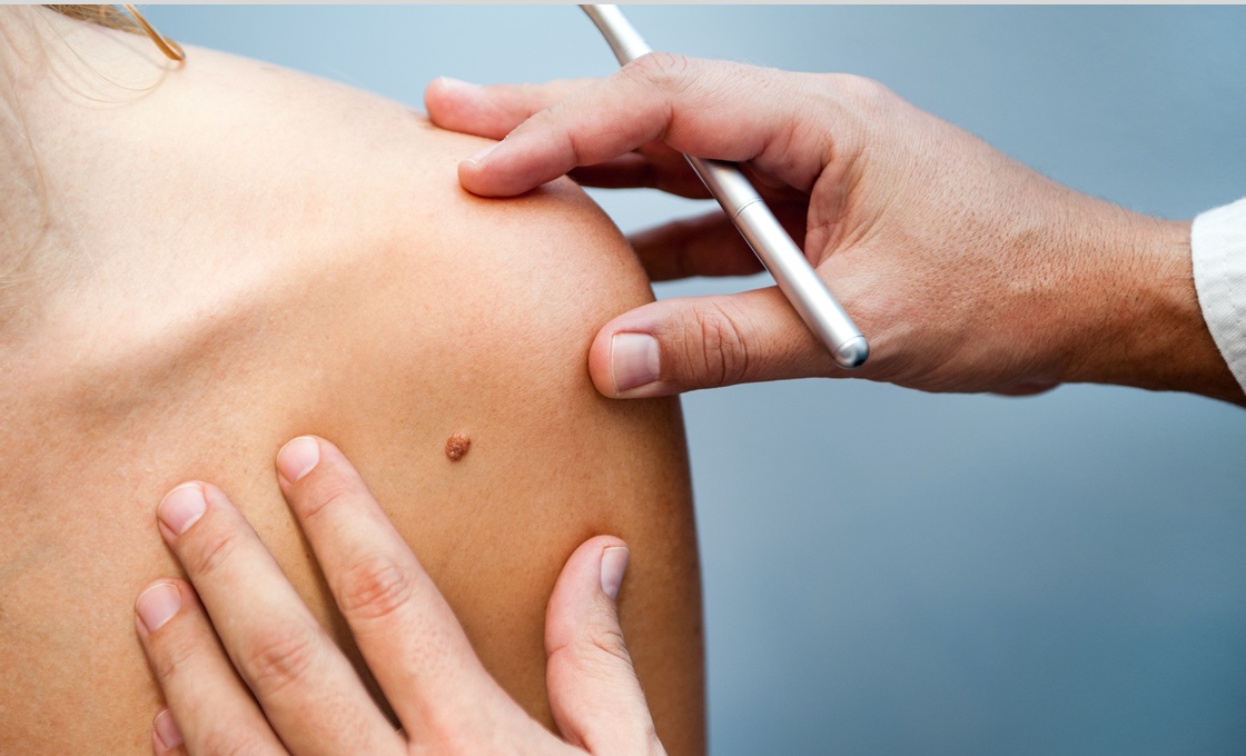
A skin self-exam will help you become familiar with the moles and spots on your body, making it easier for you to spot any irregularities that could indicate the early signs of cancer. Our dermatologists recommend examining your skin about once a month. To perform a skin self-exam, you’ll need to carefully examine your entire body from head to toe. Use a full-length mirror to examine your arms and the front and back of your body. You should use a hand-held mirror to check your scalp, the back of your neck, and buttocks. Don’t forget to look at your hands, between your toes, and the soles of your feet.
REQUEST MOHS SURGERY AT THIS CLINIC FIND AN SRT CLINICSkin Cancer Types
Here’s a closer look at the three most common skin cancer types:
Basal cell carcinoma: This is the most common type of all skin cancers. It typically grows slowly and usually develops on areas of the body frequently exposed to the sun, such as the face, neck, and head. Basal cell carcinoma usually appears as a shiny bump or nodule, pink growth, or flat lesion that’s brown or flesh-toned.
Squamous cell carcinoma: Squamous cell carcinoma also grows slowly, but it can grow deep into the skin. It frequently develops on sun-exposed skin, including the face, ears, lips, and hands, and can appear as a scaly patch, a red bump that’s firm to the touch, or an open sore.
Melanoma: Melanoma spreads quickly and is the most serious form of skin cancer. It can develop within a mole that you already have on your skin, or it can appear suddenly as a dark spot or lesion that looks different from the rest of the marks on your skin. Melanoma can also appear as a black, blue, white, or red/pink lesion or mole with a jagged or irregular border.
Skin Cancer Symptoms
What skin cancer symptoms should you be looking for when performing a self-check of your skin at home? Any spot or mark on your skin that’s new, is different from others, or one that changes in shape, color, size, or feel. You should also pay attention to any itching, bleeding, crusting, or oozing of any spots or marks. If you notice any unusual or suspicious marks on your skin, make an appointment to see a Forefront dermatologist.
Skin Cancer Diagnosis
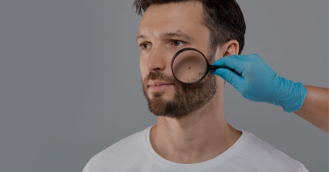
To determine a skin cancer diagnosis, your dermatologist will examine your skin, carefully looking at the lesion or spot that might be cancerous. If it looks like it could be cancerous, your doctor will remove a sample of it for further testing. This skin biopsy can determine which of the skin cancer types you have.
Skin Cancer Treatment Options
Your skin cancer treatment options depend on the size, type, and location of the lesion. Small skin cancers found on the surface of your skin may require only minor surgery, but deeper cancers typically need more extensive treatment. Treatment options for the different types of skin cancers include:
Excision: Your Forefront dermatologist will cut away and remove the growth, closing the incision with stitches.
Cryotherapy: This treatment option uses liquid nitrogen to freeze and destroy the cancerous tissue.
Curettage and electrocautery: Once your dermatologist scrapes away the layers of cancerous tissue, it’s cauterized with an electric needle. This destroys any remaining cancer cells.
Photodynamic therapy: During this procedure, a combination of laser light and special chemicals is used to kill cancer cells.
REQUEST AN APPOINTMENTMohs Surgery
Also known as Mohs micrographic surgery, Mohs surgery is regarded as the most effective treatment for high-risk, nonmelanoma skin cancers including squamous cell carcinoma and basal cell carcinoma, the two most common forms of skin cancer. Your Forefront dermatologist might recommend Mohs surgery for skin cancer if your skin cancer returns after receiving previous treatment. It’s also especially useful in treating skin cancers that have uneven edges, are large, aggressive or growing rapidly, or appear on a part of your body where it’s important to preserve healthy tissue, such as your scalp, nose, eyes, lips, ears, toes, fingers, or genitals.
REQUEST MOHS SURGERY AT THIS CLINICMohs surgery skin cancer treatment is performed in stages during a single visit to the office. Your Mohs surgeon uses a scalpel to remove the visible tumor and a thin layer of the surrounding skin underneath. The area is bandaged so you can wait comfortably while the removed tissue is examined under a microscope. In most cases, you won’t leave your appointment until all of the skin cancer has been removed.
Your Forefront Mohs surgeon may schedule a follow-up appointment with you to monitor your recovery process, make sure your wound is healing properly, and answer any questions you may have.
Superficial Radiation Therapy (SRT)
This non-surgical method uses radiation to deliver electromagnetic energy to target and destroy cancerous cells. It only penetrates the surface of your skin, minimizing scarring and preserving surrounding healthy skin tissue.
Superficial Radiation Therapy (SRT) available at these locations
1 Location in the Thousand Oaks, CA area
Interested in Skin Cancer Treatment? Request a consultation with a skin specialist today.
*Treatment options may vary at each location.Please confirm your desired treatment is offered at your preferred location when scheduling. *Age Restriction.
For patients scheduling who are under 18 years of age (19 in Alabama) please make sure you have permission from your parent or legal guardian to schedule this appointment. Your parent or legal guardian must accompany you on your initial visit and on certain subsequent visits to provide appropriate informed consent.